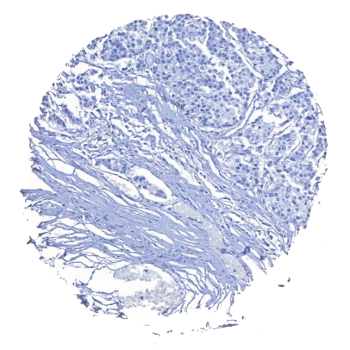
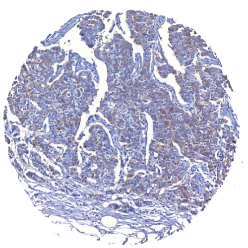
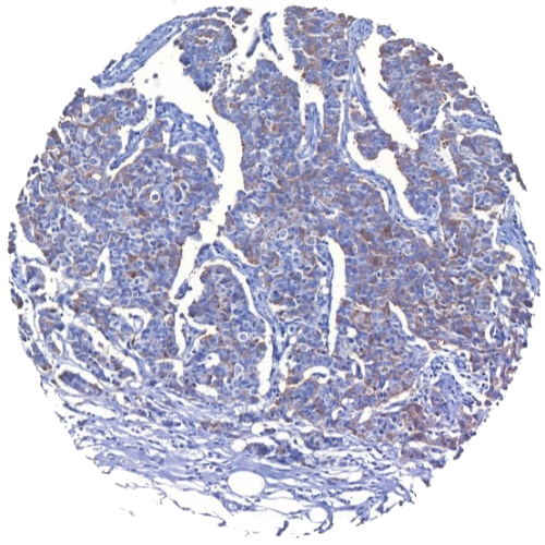
Breast Tumor Tissue Microarray
A breast tumor tissue microarray (TMA) is a cutting-edge tool designed to enhance the efficiency and accuracy of breast cancer research. By integrating multiple tissue samples into a single paraffin block, TMAs allow researchers to conduct high-throughput analysis with unmatched convenience. This technology plays a pivotal role in understanding breast cancer biology, enabling the study of biomarkers, breast tumor heterogeneity, and development of subtype-specific therapies.
Our Breast Tumor TMAs
MYmAb Biologics provides breast tumor tissue microarrays from Southeast Asian (SEA) samples globally. All our breast tumor TMAs are IRB-approved and fully consented. If you are looking for breast tumor TMA slides for your cancer research, visit our product pages or contact our team for a quote.
-
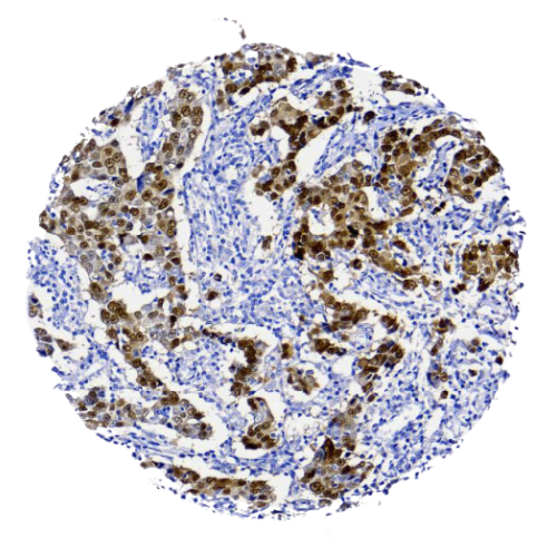
Human Tissue Microarray | Breast Tumor & Adjacent Normal Tissue Microarray – MB006
Read more -
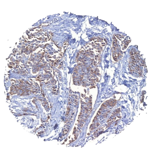
Human Tissue Microarray | Breast Tumor Tissue Microarray (120 cases) – MBB4
Read more -
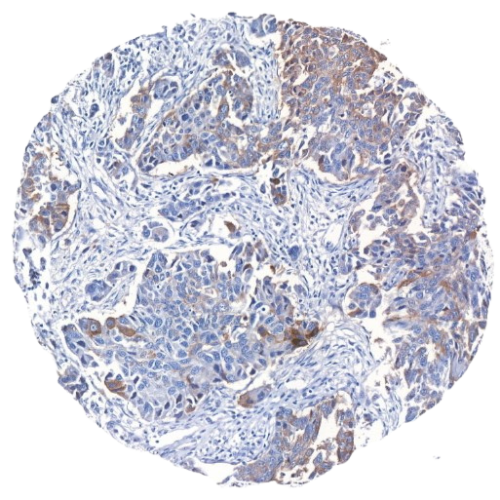
Human Tissue Microarray | Breast Tumor Tissue Microarray (129 cases) – MBB1
Read more -
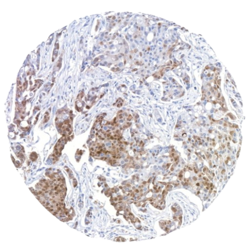
Human Tissue Microarray | Breast Tumor Tissue Microarray (131 cases) – MBB3
Read more -
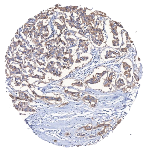
Human Tissue Microarray | Breast Tumor Tissue Microarray (88 cases) – MBB2
Read more
Our Breast Cancer Subtype TMAs
MYmAb Biologics also provides breast cancer subtype tissue microarrays from Southeast Asian (SEA) samples globally. All our breast cancer subtype TMAs are IRB-approved and fully consented to. If you are looking for breast cancer TMA slides for your cancer research, visit our product pages or contact our team for a quote.
Benefits of Breast Tumor TMAs
Breast tumor tissue microarrays offer a powerful and efficient solution for cancer researchers, enabling the analysis of multiple tumor samples simultaneously. This approach streamlines workflows, reduces costs, and ensures consistent, high-quality data, making it an indispensable tool for advancing breast cancer studies.
- Efficiency: Evaluate hundreds of tumor samples in parallel, saving time and resources.
- Cost-Effectiveness: Reduce reagent and slide preparation costs by consolidating sample analysis.
- Reproducibility: Standardized arrays ensure consistency across experiments.
- Comprehensive Data: Study a wide range of tumor stages, subtypes, and patient demographics in one slide.
- Customizability: Tailored TMA solutions to meet specific research requirements.
Applications of Breast Tumor TMAs in Cancer Research
- Biomarker Discovery: Identify and validate biomarkers for breast cancer diagnosis, prognosis, and treatment.
- Drug Development: Evaluate drug efficacy and predict therapeutic responses using diverse tumor samples.
- Histological Studies: Explore tumor morphology and progression across a wide spectrum of cases.
- Genomic and Proteomic Analysis: Perform high-throughput molecular studies to uncover genetic and protein-level changes in breast cancer.
- Personalized Medicine Research: Support research efforts toward tailored cancer therapies based on individual patient profiles.
Why Choose Our Breast Tumor Tissue Microarray
At MYmAb Biologics, we are dedicated to advancing life sciences through high-quality, ethically sourced human tissue microarrays (TMAs) tailored for global research needs. Specializing in Southeast Asian (SEA) samples, we provide researchers worldwide with IRB-approved and fully consented TMAs. Our collection spans diverse human tissues—such as breast, liver, and colorectal—covering normal, adjacent normal, and tumor samples to support comprehensive cancer studies.
Our mission is to empower scientific breakthroughs, accelerating research from the lab to clinical application. We envision a world where our contributions help extend the lifespan and enhance the quality of life for cancer patients everywhere.
Frequently Asked Questions
Conventionally, immunohistochemical staining has been performed on whole tissue sections. However, this traditional method, which requires the processing and staining of hundreds or even thousands of slides, is time-consuming and expensive. In contrast, a TMA allows simultaneous analysis of hundreds of tissues in one go using identical conditions. Thus, TMA technology markedly conserves reagents, saves time and greatly decreases the amount of archival tissue required for a particular study, thus preserving the tissues for other diagnostic and/or research needs.
A core size of 0.6 to 2.0 mm is adequate to represent the whole tissue section.
Sometimes, a small core of sample may not be representative of the whole tumour, especially in heterogenous tumour types. To overcome this, we offer TMAs that include two to three cores of samples from different locations of a single tumour block.
Negative and positive control TMA slides should be included as they are important in ensuring the validity of the experiment.
To help with TMA orientation.
A minimum of 3 slides per TMA type to include positive and negative controls.
All patients are anonymised.
Yes. You may contact us for further details and discuss on your needs.
The TMAs are sent in ambient temperature.
We recommend you to store your TMA slides at 4°C for not more than a year.
Request for Quote
Location:
Block D-G-8, UPM-MTDC Technology Centre III, Universiti Putra Malaysia, 43400 Serdang, Selangor Darul Ehsan, Malaysia.
Phone:
03-8938 9819
Email:
info.mymab@gmail.com
