数字病理学 – 保护研究成果的先进方法
数字病理学为当今医学和科学领域带来了突破,是病理学发展中的一个显著创新。它彻底改变了病理学家和研究人员的病理诊断和数据保存介质。无论您是在进行癌症研究、临床试验,还是任何组织病理学研究,数字病理学都在改变我们阅片分析、存储和共享切片的模式。此文将探讨数字病理学的基本原理、其应用范围,以及它如何成为保护和提升研究成果的关键。
什么是数字病理学?
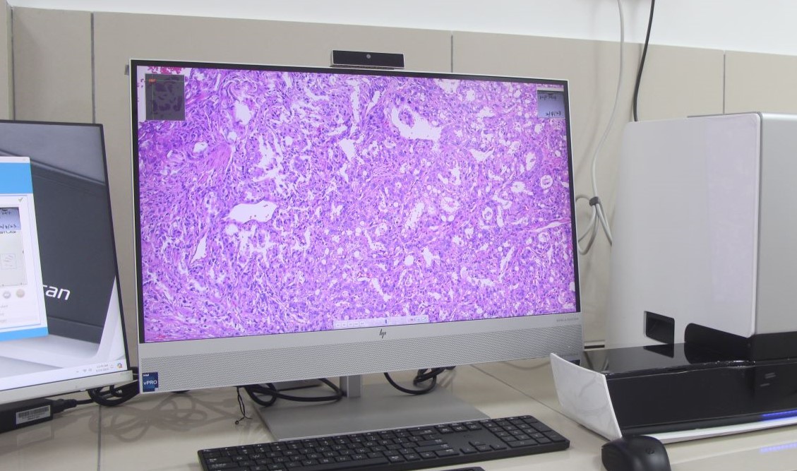
数字病理学 (Digital Pathology) 的核心是将装有生物组织样本的玻片进行数字化处理。这一过程涉及将玻片扫描为高分辨率的数字图像。这些图像可以被分析、编辑,存储并以互联网传输方式共享,为病理研究带来了极大的灵活性和可及性。
数字病理学公司利用先进的扫描仪和软件将玻片以数字化呈现,保证了准确性和便捷性。 病理切片扫描服务 可以显著优化工作流程,减少手动操作,并为样本创建可永久保存的数字图像。
病理学数字化的目的是什么?
数字病理学的兴起始于图像处理技术公司开始为病理学 开发高分辨率扫描仪。自那时起,持续的创新与突破使数字病理学成为医学研究和临床诊断中的主流实践。
病理学数字化的驱动力是对效率、精确性和远距离协作的需求。传统上,病理学家和研究人员严重依赖实体玻片,这些切片可能会随时间褪色、受损或在远程位置难以获取。而数字化病理旨在通过以下方式克服这些局限性:
- 促进协作:数字化图像使病理学家和研究人员能够轻松地共享切片,并实现全球范围的实时协作。
- 数据保存:高分辨率的切片图像可以保留样本的质量,实现长期存档,无需担心实体切片的物理退化。
- 提高效率:自动扫描仪和软件驱动的病理分析提升了诊断和研究成果的速度。
传统病理学与数字病理学的区别是什么?
在传统病理学中,病理学家使用显微镜观察染色组织样本(如H&E染色或IHC染色)的实体切片,这需要进行实体操作和现场分析。 而数字病理学则使用全切片图像(Whole Slide Image)或其他图像模式,将相同的染色组织切片捕捉为高分辨率的数字图像。这些图像可以在计算机上查看,通过网络传输与同事共享,或使用计算机辅助病理诊断。
数字病理学相较于传统病理学的优势
- 远程访问:通过数字病理学,您无需亲临实验室即可查看切片。数字切片可以通过互联网传输随时随地访问,促进远程病理诊断或者协商诊断。
- 数据存储与安全:传统切片易受到物理损坏,例如褪色或破损。数字病理学通过数字格式存储样本高清图像,避免了物理退化的风险。
- 提高准确性:数字病理系统可整合人工智能和机器学习算法进行自动图像分析,可提升人工诊断的准确性并减少人为错误。
数字病理学在转化研究中的应用
数字病理学在 转化研究(将基础科研成果转化为临床应用的过程)中发挥着日益重要的作用。以下是其支持研究人员的几种方式:
- 生物标志物发现:研究人员可以通过分析切片图像,识别潜在的疾病生物标志物,加速新诊断工具和治疗方法的开发。
- 癌症研究:数字病理学广泛应用于癌症研究,实现了精确的肿瘤分析、定量化以及细胞变化的追踪。
- 药物研发制药公司常利用数字病理学评估临床前研究中的组织样本,从而更好地了解药物与特定组织的相互作用。
- 临床试验:数字病理学可以高效准确的诊断组织样本,提供了能加速临床试验进程的关键数据。
结论
综上所述, 数字病理学 为管理和分析研究成果提供了一种创新、高效且可靠的方法。从增强协作和准确性到长期保存,其优势显而易见。
在 MYmAb Biologics我们深知精确性和速度在研究中的重要性。因此,我们为全球研究人员提供高分辨率且价格实惠的 病理切片扫描服务。 我们的尖端技术确保您的切片以最高质量进行数字化处理,让您专注于最重要的工作——您的研究。不论您从事的是癌症研究还是其他组织病理学研究,我们随时准备为您服务。
请立即联系 我们 以了解我们的数字病理学服务如何助力您的研究迈向新高度!